Investigational AI feature
This AI radiograph-analysis feature is investigational and not cleared, approved, certified, or registered for diagnostic use. Outputs are candidate observations only and must be independently reviewed, accepted, rejected, or edited by a qualified clinician. Do not use as the sole basis for diagnosis, treatment planning, referral, prescribing, insurance claims, or patient communication.
AI-assisted radiograph review support
Candidate observations for clinician review.
Cavitech AI Vision may highlight candidate radiographic observations across panoramics, periapicals, and full mouth X-rays while the clinician remains responsible for review and sign-off.
Cavitech AI VisionLearn more
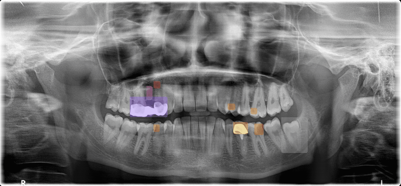
Candidate observation
Anatomy marker
Bone segmentation
Candidate observationTooth 36
AI 2DClinician review85%
Accept
Reject
Edit
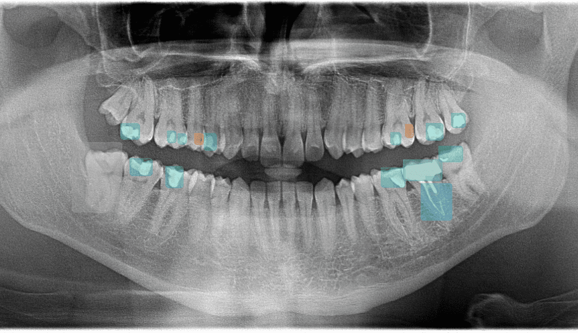
Candidate observations and anatomy markersClinician review required
28observation types
<10sAverage evaluation run time
Supported Formats
Every X-ray your practice takes.
Panoramic (OPG)Bone levels, impacted teeth, sinuses, and broad candidate-observation review across all quadrants
Periapical (PA)Candidate periapical, root, caries-depth, and endodontic observations at tooth level
3D Imaging (CBCT) – (Coming Soon)3D CBCT review support, implant planning, nerve tracing, and volumetric candidate observations - coming soon
Candidate observations
28 observation types.
4 clinician-review categories.
Restorative · 7
CariesSecondary CariesCrown FractureDefective RestorationMissing ToothOverhangOpen Margin
Periodontal · 6
Bone LossCalculusFurcation InvolvementWidened PDL SpaceVertical Bone DefectHorizontal Bone Loss
Endodontic · 8
Periapical LesionRoot ResorptionRoot FractureShort Root Canal FillingOverfilled CanalPeriapical AbscessInternal ResorptionPulp Stone
Anatomical & Other · 7
Impacted ToothSupernumeraryRetained RootSinus PathologyIAN Canal ProximityCystAbnormal Morphology
Review support layers
Context for clinician-controlled radiograph review.
01Anatomy DetectionMay mark IAN canal, mental foramen, maxillary sinus boundaries, and root apices for clinician review
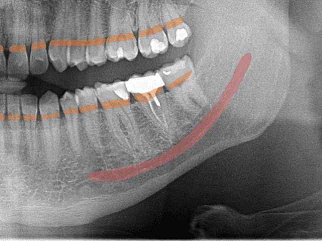
Landmarks
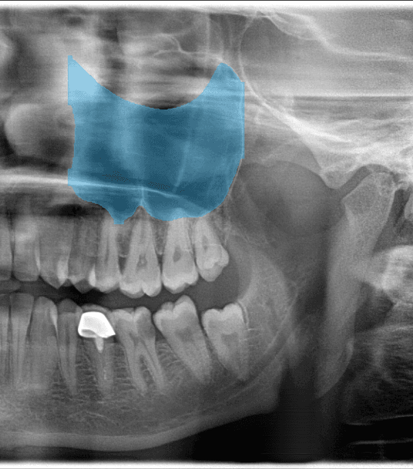
02Bone SegmentationMay support clinician review of bone levels and possible horizontal or vertical defect patterns
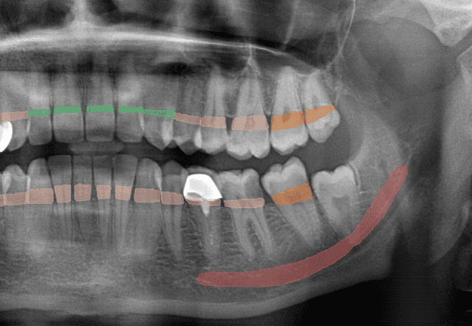
Bone Levels
03Sinus DetectionMay highlight sinus boundaries and possible patterns for clinician review in planning context
Sinus
04Teeth Detection & FDIMay number visible teeth using FDI notation (11-48) and associate candidate observations with positions
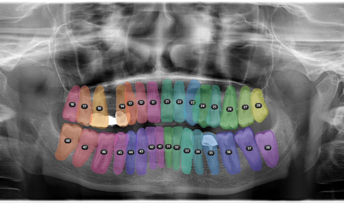
FDI Map
05Soft Tissue InsightsMay highlight candidate calcification, asymmetry, or soft-tissue-region observations for clinician review
06Enamel HealthMay flag candidate demineralisation, erosion, and defect patterns for clinician review
07Decay ProgressionTrack caries across visits over time — visualise changes in depth and severity
Get Started
Evaluate AI-assisted radiograph review support.
Invite only - investigational evaluation - panoramic, periapical, and FMX