AI Diagnostics
28 pathologies. 3 X-ray types. Seconds, not minutes.
Cavitech AI Vision analyses panoramics, periapicals, and full mouth X-rays — detecting pathology, mapping anatomy, and numbering every tooth automatically.
Cavitech AI VisionJoin the BetaLearn more
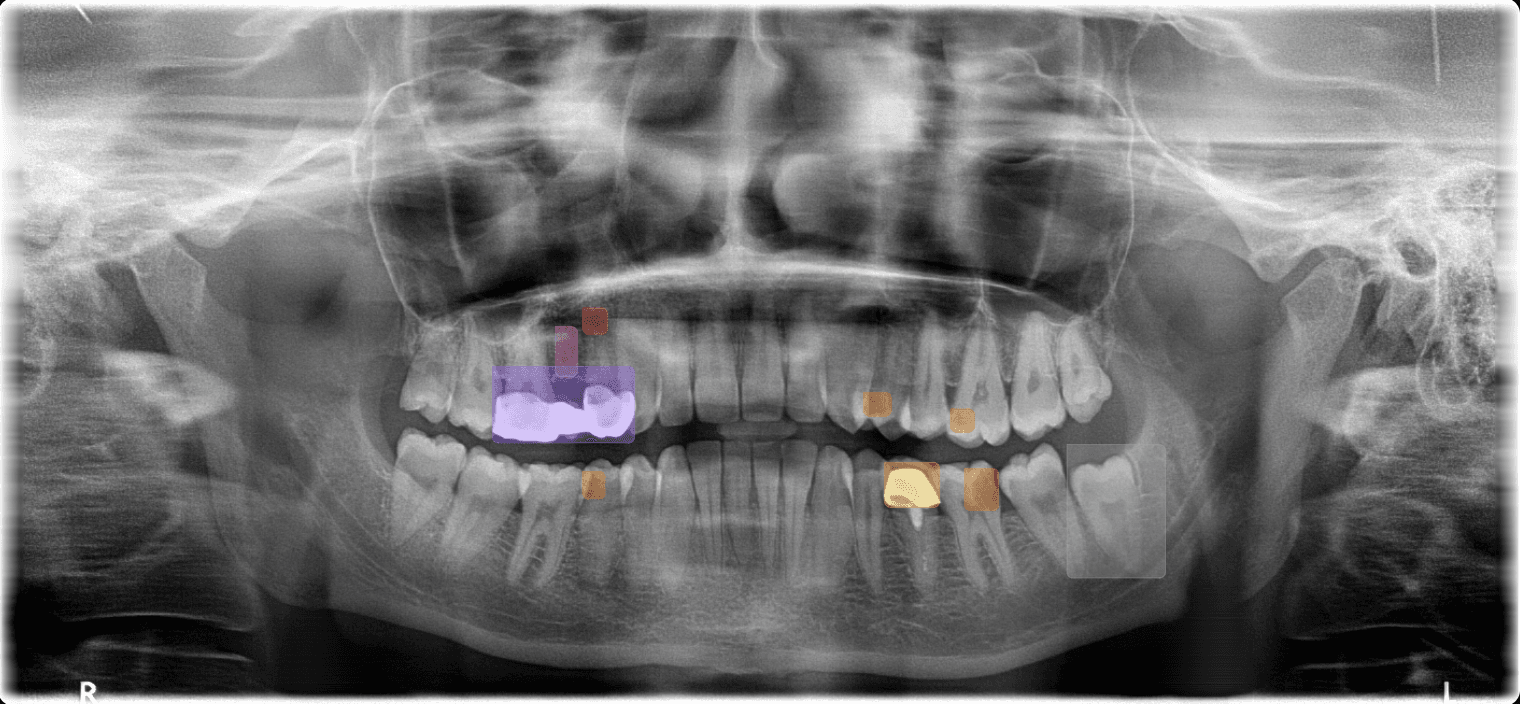
Pathology Detection
Anatomy Detection
Bone Segmentation
CariesTooth 36
AI 2DPendingModerate85%
✓ Confirm
✕ Decline
Pathology & Anatomy DetectionCaries & 28 other
28Pathologies
<10sAverage analysis time
Supported Formats
Every X-ray your practice takes.
Panoramic (OPG)Bone levels, impacted teeth, sinuses, and broad pathology screening across all quadrants
Periapical (PA)Periapical lesions, root fractures, caries depth, and endodontic assessment at tooth level
3D Imaging (CBCT) – (Coming Soon)3D CBCT analysis, implant planning, nerve tracing, and volumetric pathology projection — coming soon
What We Detect
28 pathology types.
4 clinical categories.
Restorative · 7
CariesSecondary CariesCrown FractureDefective RestorationMissing ToothOverhangOpen Margin
Periodontal · 6
Bone LossCalculusFurcation InvolvementWidened PDL SpaceVertical Bone DefectHorizontal Bone Loss
Endodontic · 8
Periapical LesionRoot ResorptionRoot FractureShort Root Canal FillingOverfilled CanalPeriapical AbscessInternal ResorptionPulp Stone
Anatomical & Other · 7
Impacted ToothSupernumeraryRetained RootSinus PathologyIAN Canal ProximityCystAbnormal Morphology
Annotated X-ray
Beyond Pathology
Every layer of the radiograph.
01Anatomy DetectionMaps IAN canal, mental foramen, maxillary sinus boundaries, and root apices automatically
Screenshot
Landmarks
02Bone SegmentationMeasures bone levels around each tooth — identifies horizontal and vertical defects with precision
Screenshot
Bone Levels
03Sinus DetectionIdentifies sinus boundaries, mucosal thickening, and pathology relevant to implant planning
Screenshot
Sinus
04Teeth Detection & FDINumbers every visible tooth using FDI notation (11–48) and links all findings to positions automatically
Screenshot
FDI Map
05Soft Tissue InsightsDetects calcifications, asymmetries, and abnormalities in visible soft tissue regions on radiographs
Screenshot
06Enamel HealthFlags early demineralisation, erosion, and defects
07Decay ProgressionTrack caries across visits over time — visualise changes in depth and severity
Get Started
See what your X-rays are missing.
Free beta · 28 pathology types · Panoramic, periapical, and FMX